Tag Archive for: body systems

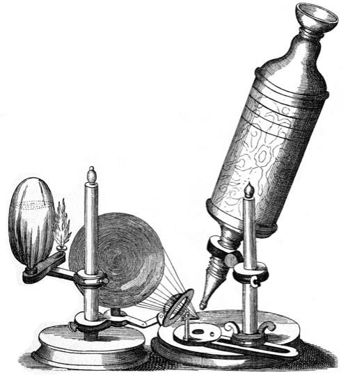
The year is 1665. The Taj Mahal in India was completed 12 years ago. In a little over a year, Isaac Newton will witness an apple falling from a tree, sparking an idea. And somewhere in London, the architect and natural philosopher Robert Hooke places a thin slice of cork into the specimen holder of a microscope. When he looks through the eyepiece, he sees a strange structure.

“I could exceedingly plainly perceive it to be all perforated and porous, much like a honeycomb, but that the pores of it were not regular,” he writes. “These pores, or cells … were indeed the first microscopical pores I ever saw, and perhaps, that were ever seen, for I had not met with any Writer or Person, that had made any mention of them before this.”

Hooke has discovered the cell. Plant cells to be specific. He actually coins the term, writing that they remind him of the cells occupied by Christian monks in a monastery he once visited. These cells are dead though, and his microscope is not powerful enough to see inside the cell. It’s not until 13 years later that someone would see a living cell up close.

Using a more powerful microscope of his own design, Dutch businessman and scientist Antonie van Leeuwenhoek would first observe bacteria and protozoa. He called these single celled organisms animalcules, Latin for “little animals.”

Hooke is long gone now, buried somewhere in the City of London Cemetery. He took the first steps towards what is now refer to as cell theory. This is the understanding that every living organism on the planet is composed of one or more cells.

Cells are the integral unit of structure and function in all living organisms. Every cell that has ever existed came from pre-existing cells that have divided, and divided, and divided, all the way to the 37.2 trillion cells that make your body.

The Two Different Types of Cells

Cells can be split into two main types—prokaryotes and eukaryotes.

Prokaryotic cells do not have a nucleus. Those “little animals” that Leeuwenhoek witnessed were prokaryotic cells. Bacteria, and another family of cell called archaea, are classified as prokaryotic.

The cells that exist in plants and animals are called eukaryotes. This type can be either single-celled or multicellular.

Approaching the Cell

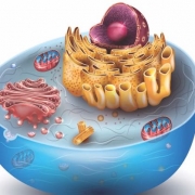
But what makes up a eukaryotic animal cell? If you could shrink down to the size of the cell, and even smaller, what would you see?

Imagine you’re getting smaller and smaller. The world around you gets larger and larger, eventually blurring out of view. As you shrink, you start to focus in on a group of structures, like the little cages that Hooke witnessed long ago.

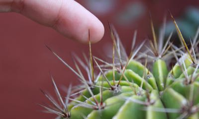
Soon enough you come to one cell in particular. Now, some cells are more complex on the outside and have accessories other cells lack. Microvilli are one such feature.

Microvilli extend like fingers from the surface of the cell, and are important in the absorption of nutrients. They also greatly increase the surface area of the cell without affecting its overall size.

Cilia extend even further than microvilli, and can actually push different substances along the surface of the cell.

Then there is the flagellum, which is a thin, tail-like structure that can actually propel an entire cell, enabling it to swim!

The Plasma Membrane

All cells rely on the all-important plasma membrane. This acts like a fence, keeping the contents of the cell together while also letting food and nutrients pass through.

The plasma membrane is made up of a double layer of fatty acids called phospholipids. These fatty acid molecules have a head and a tail. The head is what is called ‘hydrophilic,’ meaning it’s attracted to water. The tail, meanwhile, is hydrophobic—repelled by water. This combination of head and tail is what makes the structure and function of the cell membrane possible.

As you get smaller, you pass through the plasma membrane, and journey into the cell. Briefly, you can see the double layer of phospholipids, like a zipper held fast by the chemical attractions of their hydrophobic tails.

Cytoplasm and Cytoskeleton

Once fully inside the cell, you encounter a medium called the cytoplasm. It contains a substance rich in amino acids and potassium, called cytosol. This solution is also referred to as intracellular fluid.

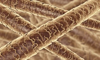
You can also make out a network of what looks like webs or scaffolding. This is the cytoskeleton. It provides structural support and allows the movement of materials inside the cell. The cytoskeleton is made up of three different types of protein fibers called microfilaments, intermediate filaments, and microtubules.

Microfilaments are the smallest of the three, made of twisted strands of proteins that can be pulled together to shorten the cell. This occurs often in muscle cells, and aids in their ability to contract.

Intermediate filaments are twisted strands of proteins that mainly provide framework for the cell and help hold it together.

Microtubules have a spiral shape. When put together, they form a hollow cylinder. These cylinders help maintain cell shape and move organelles (another name for cell parts) within the cell.

They form what is called the centrosome. The centrosome is made up of structures called centrioles which organize microtubules and provide an additional framework for the cell. They also aid in the separation process during cell division.

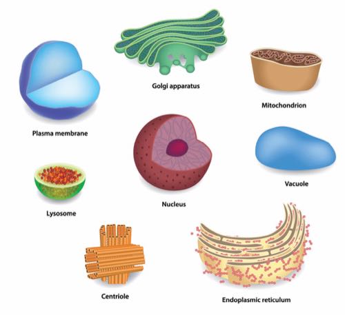
Between the cytoplasm and the cytoskeleton, you can see the primary support framework of the cell. You can also see several strange-looking structures. These are the organelles. These important cell parts all have specific functions they carry out.

The Endoplasmic Reticulum

The first structure you can see looks like a collection of several long, thin caverns. These are the endoplasmic reticulum (ER). There two different types of ERs.

The first is the rough ER, which extends from the nucleus and has ribosomes attached to the outside of its membrane, giving it a rough appearance. These ribosomes produce what are called polypeptide chains. That’s just a fancy way to say proteins. The proteins created by ribosomes are released into the ER, where they are processed and prepared for release into the cell. When released, the proteins are transported inside enclosed membrane sacks called transport vesicles that pinch off from the rough ER.

It’s important to note that ribosomes are not organelles. They are vital to cells, though. That’s because they’re the protein-producing factories. They can either be floating in cytosol en route to somewhere else in the cell, or attached to the rough ER. Ribosomes are comprised of two components called the small and large subunits. The small subunits read the ribonucleic acid (RNA), which contain instructions on how to assemble the amino acids into polypeptide chains. The large subunit does the heavy lifting of actually assembling the polypeptide chains.

Next you see the smooth ER. This is another organelle with a membrane, but it doesn’t have ribosomes, hence the “smooth” moniker. The smooth ER contains enzymes that alter polypeptides, produce lipids and carbohydrates, and destroy toxins. Most of the lipids and cholesterol that make up cell membranes are made in the smooth ER.

The Golgi Apparatus

Shifting your focus, you encounter the Golgi apparatus, definitely the coolest name of all the organelles. The Golgi apparatus is another membranous organelle that modifies, packages, and stores proteins.

It looks like a group of larger and larger cisterns expanding out from its center. Transport vesicles deliver proteins to the Golgi apparatus from the ER. As the proteins move throughout the cisterns of the Golgi, they are modified. This can happen by adding or rearranging molecules with different enzymes. Sometimes carbohydrates are added to form what are called glycoproteins.

After moving through the last cistern, proteins are cordoned off in a different vesicle called the secretory vesicle. Most of these proteins are directed toward the plasma membrane. They either become part of the membrane, or are released outside of the cell.

Lysosomes

The Golgi is fundamental in the production of lysosomes. These are vesicles that pinch off from the Golgi apparatus and function as the garbage trucks of the cell. Lysosomes are enclosed by a membrane and contain digestive enzymes that pick up cellular waste or defective organelles to be recycled or converted to waste. They are also vital in protecting the cell from bacteria and viruses.

Proteasomes

Passing out of the Golgi apparatus, you come across the proteasomes. These organelles manage the existing proteins in the cell. They are found throughout the cytoplasm. Proteasomes break down abnormal or misfolded proteins and normal proteins the cell doesn’t need anymore.

Another protein called ubiquitin is placed on the proteins marked for recycling by enzymes in the cytoplasm. The targeted proteins are then pulled into the proteasomes and broken down by a process called proteolysis. In this process, the peptide bonds of the proteins are broken. The leftover peptide chains and amino acids are then released into the cell to be recycled.

Peroxisomes

Moving on, you come across a curious structure called a peroxisome. While not technically an organelle, and not technically an enzyme, peroxisomes can best be described as protein complexes.

They have a membrane, and are also pinched off from the ER. Peroxisomes are responsible for breaking down long-chain fatty acids and amino acids. In this process, they can produce the byproduct hydrogen peroxide, which can be dangerous to the cell because it can react with many substances. Because of this, peroxisomes also carry an enzyme that converts hydrogen peroxide into water and oxygen. Talk about cleaning up after yourself!
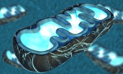
Mitochondria

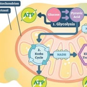
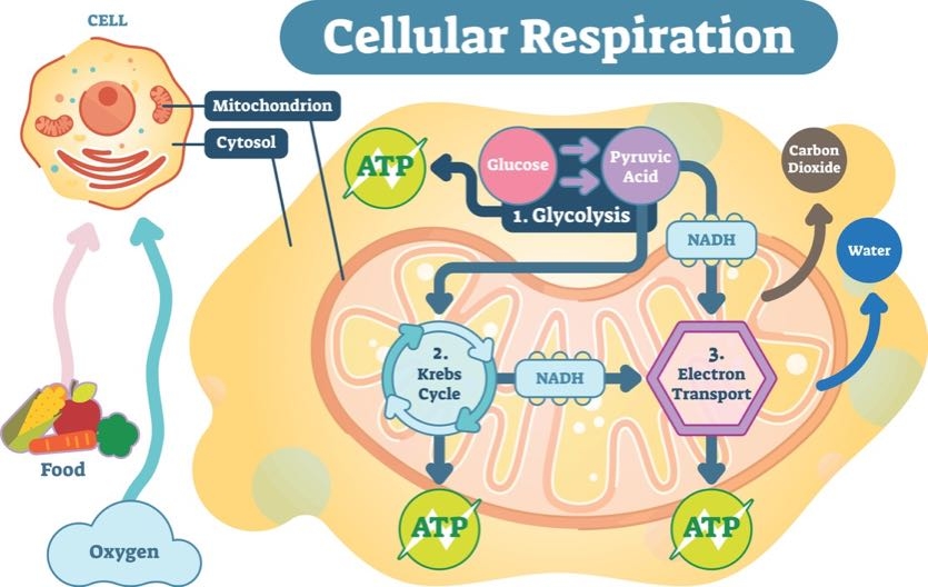
Once past the peroxisomes, you spot a baked-bean-shaped organelle called a mitochondrion (when there are many, they’re called mitochondria). These are the hyper-efficient power plants of the cell. They take food particles brought into the cell and convert it to a molecule called adenosine triphosphate, or ATP. This is known as the “currency” of the cell. ATP is capable of storing and transferring energy to other parts of the cell.

Mitochondria have both an inner and outer membrane, and their numbers can vary depending on the type of cell. Typically, the more active a cell is, the more mitochondrion it will contain. Liver cells, for example, contain thousands of mitochondria. In the cells that make up your muscles, aerobic activity can actually increase the number of mitochondria. No wonder you have more energy when you exercise frequently.
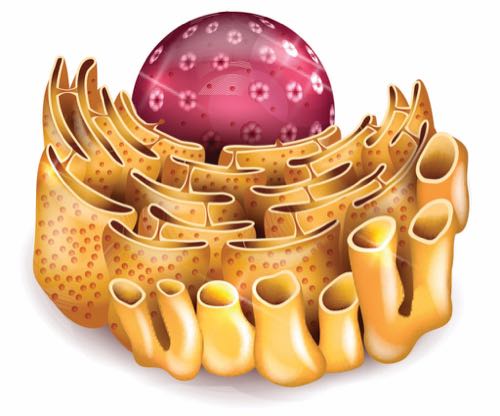
The Nucleus

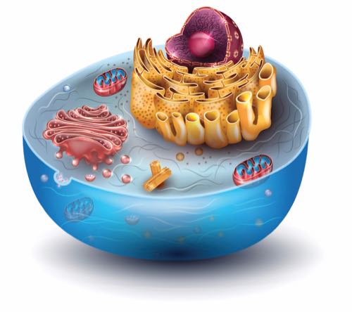
Finally, you arrive at the nucleus. The largest of all the structures in the cell, the nucleus has two membranes forming what is called the nuclear envelope.

Along with small pores on the surface of the membrane, this envelope encloses the nucleoplasm. While the nuclear envelope functions as a wall, the pores act as a gate that lets certain molecules in and out of the nucleus. Nucleoplasm is similar to the cytoplasm of the cell. It is a syrupy substance that suspends the structures contained within the nuclear membrane.

Suspended in the nucleoplasm is the nucleolus. It is comprised of deoxyribonucleic acid (DNA), RNA, and protein. The nucleolus is the birthplace of ribosomes, which, remember, make proteins vital to the functioning of healthy cells.

As you get smaller, you can start to make out the twisted double-helix structure of the cell’s DNA. You reach out, trying to touch it, closer and closer, smaller and smaller. And finally, you make contact. In a flash, you return to your previous size, not sure whether or not you actually touched what you were reaching for.

Somewhere in the grassy fields of the City of London cemetery, the first light of a brand-new day strikes a freshly germinated seed of grass. The cells of that seed, enriched by the good earth and sun, divide and divide, sending forth a tiny shoot into the cool morning air.

You use your teeth every day, but you might not know much about their anatomy. It’s time to start asking questions and learning more about them. Maybe “what are teeth made of?” has popped into your mind before. Or you’ve wondered from time to time: are your teeth bones?

It’s your lucky day.

You’ve found 24 bite-sized questions and answers to help you better understand your teeth. From baby teeth to teeth grinding, plaque, and flossing, this list has got it all. Study up so you can display your knowledge the next time you’re at the dentist.

  1. What Keeps Your Teeth in Place?

Your teeth are anchored into two of the bones of your skull. The upper teeth are situated in a bone called the maxilla that forms the upper jaw. And the mandible (jawbone) is the bone that houses your lower teeth.

The mandible and maxilla are the two bones that make up your jaw. They are connected on the right and left sides of your skull. Your jawbone is the strongest in your skull and the only one that can move. And that comes in handy when you bite and chew your food.

  1. How Many Teeth Do You Have?

Ancient philosophers like Aristotle once thought men and women didn’t have the same number of teeth. But we know better now. A full set of adult teeth for women and men is 32. You can count them yourself with your tongue.

Teeth are organized in pairs according to their shape. You have one tooth from the pair on each side of your mouth. Look in a mirror and see for yourself. Divide your mouth in half vertically to see that each side is virtually symmetrical.

  1. What is Your Tooth’s Crown? (Not the One from the Dentists, Either)

To answer this question, let’s breakdown the anatomy of a tooth.

Teeth are like icebergs. The iceberg appears to be a small chunk of ice floating in the water. When really, it’s like an ice mountain barely peeking up out of the ocean. So it is with your teeth. They exist above and below the surface of your gums. The shiny, white portion you can see when you open your mouth is called the crown.

This part of the tooth comes in contact with your food, drink, and saliva. It is the tough exterior that protects the whole tooth. So, a tooth’s crown is covered in a hard substance called enamel. Underneath the enamel is a layer of hard tissue called dentin.

  1. What is Enamel?

Until now, you probably thought that bone was the toughest tissue in your body.

Wrong!

That award goes to enamel. The hard, white material that surrounds the crown of your teeth takes the title.

Enamel is found on the outside of the tooth and made primarily of calcium phosphate. This mineral makes enamel incredibly strong. So, it can defend the softer, more sensitive layers of the tooth—dentin, pulp, nerves, and blood vessels. It also protects your teeth from the wear-and-tear of daily use.

  1. Do Teeth Have Roots?

They sure do. Below the hard crown is the tooth’s root. It’s tucked away underneath the gum line and tethered to the jawbone by connective tissue called the periodontal ligament.

Most of the root is made of dentin, which forms canals. They hold a living tissue called pulp that’s full of blood vessels and nerves that run through the root of the tooth and into the jawbone.

  1. Can Teeth Feel Sensations?

You might have noticed your teeth feeling sensitive to hot or cold temperatures. This can happen if the root of a tooth is exposed above the gum line, or if enamel wears down. When the dentin that makes up the root is uncovered, the nerves inside can be stimulated by the temperature of the food and drink in your mouth.

The sensations of hot and cold are transmitted through the nerves in the exposed dentin to the brain. Your brain interprets these signals as pain.

This is another reason enamel is so important. It acts like an insulator that shields teeth from extremely hot and cold temperatures. Enamel protects the sensitive nerves in dentin from painful stimulation.

  1. Why Do Teeth Come in Different Shapes?

Take one look at your smile and you’ll know your teeth don’t all look the same. In fact, your teeth vary widely in their shape and size.

While none of the teeth in your mouth are identical, they can be classified by their overall shape. An adult set of teeth has eight incisors, four canines, eight premolars, and 12 molars.

Beginning in the middle and branching out left and right are your incisors. These big front teeth are sharp like a knife. Next to the incisors are the canine teeth. They have a distinctive point called a cusp. They resemble the pointed teeth in dogs.

The next teeth in line are called the premolars. These teeth have two cusps and are sometimes referred to as bicuspids. Finally, the last class of teeth are the molars. They’re large and flat.

  1. What Do the Different Kinds of Teeth Do?

The different shape of each tooth helps it perform a specialized function while you chew your food.

Incisors are great at cutting into and holding chunks of food, like when you bite into an apple. Your incisors can also help you sense the texture of your food.

Canine teeth tear your food into smaller, more manageable pieces. You can put your canines to work by eating a piece of thick protein, like grilled chicken.

The premolars are between canines and molar in shape. Premolars help cut and tear food—much like the canine teeth do.

Molars are used for grinding food. As you chew, pieces of food are moved further back into your mouth where they’re ground up by your molars. Molars help break food into pieces you can swallow safely.

  1. What are Wisdom Teeth?

You have four wisdom teeth, which are also called third molars. They are shaped just like molars and perform the same tasks while you chew. But for some people, wisdom teeth need to be removed.

In your late teens and early twenties, wisdom teeth arrive. These are the last of your permanent teeth to erupt (another word for your teeth coming in). When wisdom teeth come to the surface, they can push against the other teeth, causing crowding and discomfort. They can even knock your other teeth out of alignment.

If your dentist believes your wisdom teeth will cause a problem, they’re often surgically removed. This procedure usually happens before the teeth erupt. Should you have your wisdom teeth removed, the number of permanent teeth in your mouth drops from 32 to 28. Don’t worry, you will get along just fine without your wisdom teeth. You might even be more comfortable.

  1. If Your Teeth are so Strong, How Can They Get Chipped?

The material that makes up most of your tooth enamel is called calcium phosphate. This mineral compound is also found in bones. It contributes to the white color of your teeth. And it’s incredibly strong and durable. But it isn’t indestructible.

Your teeth can get injured just like the rest of your body. If your tooth gets chipped or cracked, a dentist can repair the damage. But it will not heal on its own.

  1. Are Your Teeth Bones?

Even though they look a lot alike, teeth are not bones. And, surprisingly, they’re actually very different. Here’s how:

  • Teeth are stronger than bone. As you know from above, enamel is the strongest tissue in your body.
  • Bones are protected by layers of muscle and skin. Your teeth are covered only by your lips.
  • Even though teeth are made of tougher material than bones, your bones can regenerate used and damaged tissue. When it comes to enamel, once it is gone it doesn’t come back.
  • Both bones and teeth have blood vessels and nerves inside them. But unlike teeth, bones actually produce new blood cells in their bone marrow. Teeth have an inner layer similar to marrow. This is the pulp layer that is protected by tooth enamel.
  1. What is the Color of a Healthy Tooth?

Healthy teeth are bright and white. Their color comes from the calcium phosphate in enamel. The tips and edges of teeth may appear translucent or blue-tinted. This is perfectly normal.

Yellowing of teeth can indicate enamel loss. Dentin, the layer underneath enamel, is yellow. When enamel wears down and dentin is exposed, the tooth can become discolored and appear yellow.

Fortifying your enamel with calcium rich foods—like milk, yogurt, almonds, and edamame—can help restore its whiteness. You can also drink fluoridated water to support the health of your enamel. Fluoride reinforces enamel and keep the yellow dentin from being exposed.

  1. How Many Teeth Do Children Have?

Little children have 20 primary (or baby) teeth. They are eight incisors, four canines, and eight molars. These teeth are shed later to make room for larger, permanent teeth. By age three, most children have each of their primary teeth.

Baby teeth are made of the same materials as permanent teeth. Enamel covers the crown, and the root is made of dentin and pulp. Primary teeth are smaller and more spaced out than permanent teeth. This is because the bones of the face and jaw grow as the child gets older, causing teeth to spread out.

  1. When Do Teeth Start Developing?

Before you were born, your teeth were forming. Between the third and fourth month of pregnancy, cells called ameloblasts generate the enamel that forms teeth. This happens around the tooth bud (the first stage of tooth development). These buds stay below the surface of the gums until fully formed—anywhere from six months to a year after birth.

  1. Why Do Teeth Fall Out?

As a child, your teeth fall out so larger, adult teeth can take their place. This is a natural and healthy part of the lifecycle of your teeth. Most primary teeth will fall out by age 12.

Tooth loss happens when the roots of primary teeth dissolve in preparation for the arrival of permanent teeth. This process can take several weeks, and it’s best to let teeth fall out on their own. After a primary tooth is out, a new permanent tooth will erupt in the same spot.

  1. What are Cavities?

Just like their name implies, cavities are tiny holes in the enamel and dentin layers of teeth. Cavities are the result of tooth decay, which happens when bacteria invade broken or damaged teeth.

Simple sugars are the main culprits behind cavities. Soda, juice, candy, and similar foods can linger on teeth. The bacteria in your mouth can turn these simple carbs into acids that erode the enamel on your teeth. And a cavity is born.

You might notice that you have a cavity on your own. Many people experience toothache and discomfort when they have a cavity. Your tooth might be especially sensitive to temperature or ache when you eat something sweet.

Sometimes it takes a trip to the dentist to find a cavity. With x-ray imaging the dentist can see cavities between your teeth. They can also use dental equipment to look for soft spots and holes on the tooth’s surface.

Fortunately, dentists can fix cavities with a dental filling. First, the dentist removes the decayed portion of tooth with a small drill. Then they replace the missing part of the tooth with a safe material. Fillings can be made of gold, silver, porcelain, or a composite resin. After a filling, your tooth will feel much better.

  1. What is Plaque?

If your teeth feel fuzzy after eating, plaque is to blame. When you eat sugary foods, the bacteria in your mouth cling to your teeth and feast on the simple carbohydrates. These bacteria can form a sticky film on the surface of your teeth called plaque.

You can easily remove plaque. Brushing your teeth is the best way to get rid of plaque. Do it twice a day for best results. Rinse your mouth with anti-bacterial mouth wash to keep the number of bacteria in your mouth under control. And consider an oral probiotic to help support a healthy bacteria balance.

  1. What is Tartar?

Plaque that stays on teeth can harden into tartar—a tough, mineralized substance. When tartar is present, cavities can soon follow. And tartar makes it difficult to brush properly. It requires a professional dental cleaning to remove.

Plaque generally forms above the gum line, but tartar can build up above and below the gums. This can cause problems for the dentin and bone underneath your gums. That’s why it is so important to take care of your teeth to avoid tartar build up.

You can protect your teeth from tartar by brushing and flossing daily and using mouthwash. Another great way to keep tartar at bay is fortifying your enamel with fluoride. This mineral is found in most municipal water systems. It strengthens enamel and helps repair damage caused by the bacteria and acids in your mouth.

  1. Why Should You Brush Your Teeth?

For thousands of years people have been brushing their teeth. Ancient Egyptians in 5,000 B.C. used frayed twigs and egg shells to polish their teeth. Now toothbrushes with soft bristles clear away the food left behind after a meal.

Brushing your teeth is the best way to keep cavities away. You should brush your teeth twice a day with a pea-sized amount of toothpaste. Be gentle when you brush. Scrubbing hard with a toothbrush can irritate the gums and expose the sensitive dentin underneath.

Two minutes is the perfect amount of brushing time. Keep a timer or clock in your bathroom to help measure how long you brush. Switch sides often and cover the front and back of each row of teeth.

Change your toothbrush when it wears out. Every three to four months is recommended. Rinse your toothbrush with hot water after each use. And get a new toothbrush after you’ve been sick.

  1. Is Flossing Important?

Absolutely! Brushing cares for the parts you can see. But you need to clean between your teeth, too. Using dental floss loosens up food and plaque. It keeps tartar from building up in hard-to-reach areas. Flossing also cleans the parts of your teeth hidden below your gum line.

Floss every day to keep the areas between your teeth healthy and clean.

  1. How Can You Help Avoid Tooth Decay?

Brushing and flossing are the best ways to keep your teeth healthy and your enamel in good shape. But your diet can play an important role in helping avoid tooth decay.

Steer clear of soda, juice, and other sugary drinks. These are the most harmful. So, drink water instead. Simple carbohydrates and starchy foods should be limited. When you want something sweet, skip the candy. Look to fruits for their natural sweetness as an alternative. Their high fiber content stimulates saliva production and washes sugar off your teeth. Acidic foods—like citrus fruits—can erode enamel over time, so make sure to drink plenty of plain water when consuming them.

Foods rich in calcium like milk, yogurt, and cruciferous vegetables are great for your teeth. Celery and other crunchy vegetables help clean your teeth of debris. And follow the same advice for your teeth as you do for your waistline. That means choosing whole foods and nutritious meals over simple, sugary snacks.

  1. What is Bruxism?

Part of caring for your teeth is managing stress. Stress might tense you up and make you clench your fists. Some people also clench their jaw. This can lead to a condition called bruxism, or teeth grinding.

Bruxism can happen during sleep, so people who grind their teeth are often unaware of it. But grinding can lead to excessive wear on the flat portions of your teeth. It can also cause jaw soreness and headaches.

Your dentist can tell you if you grind your teeth. They might recommend wearing a mouthguard at night to keep your teeth from grinding together.

But there are other ways to fight bruxism. Try deep breathing exercises before bed. Hold a warm washcloth on your cheek just below the ear to help relax the muscles in your face and jaw. Place the tip of your tongue between your front teeth to open and relax your jaw.

If you’re stressed, open up to someone. Sharing your thoughts and feelings with a trusted friend or mental health professional can help manage your stress or anxiety. You might notice you sleep better and stop grinding your teeth.

  1. Can Your Teeth Give You Bad Breath?

Poor oral hygiene is usually the root of bad breath. When you skip brushing or flossing, the bacteria in your mouth go to town on the food lingering on your teeth. The breakdown of sugars and starches by bacteria creates some foul-smelling odors.

Sometimes the food you eat is the source of your stinky breath. Garlic and onions are notorious for leaving bad breath behind. Spicy foods can also share the blame.

Whether your bad breath is caused by bacteria or the food you ate, the best way to eliminate it is brushing and flossing. Brush twice a day to keep your breath fresh. If you eat a particularly pungent meal, gently clean your teeth afterward, too.

  1. Are Your Teeth Unique?

It may come as a surprise, but just like your DNA and fingerprints, your teeth are unique. No one has the same teeth you do. Their shape, size, and placement vary from person to person. Not even identical twins have the same set of teeth!

Keep Your Teeth Bright and Gleaming

Show your teeth some respect by making sure they’re in good shape. Keep them healthy by taking proper care of them. Brush and floss daily. Avoid sugary foods that erode your enamel. And visit the dentist regularly.

Smile, because there aren’t any other teeth in the world like yours.

About the Author

Sydney Sprouse is a freelance science writer based out of Forest Grove, Oregon. She holds a bachelor of science in human biology from Utah State University, where she worked as an undergraduate researcher and writing fellow. Sydney is a lifelong student of science and makes it her goal to translate current scientific research as effectively as possible. She writes with particular interest in human biology, health, and nutrition.

As soon as you get out of bed, your five senses are hard at work. The sunlight coming in through your window, the smell of breakfast, the sound of your alarm clock. All these moments are the product of your environment, sensory organs, and your brain.

The ability to hear, touch, see, taste, and smell is hard-wired into your body. And these five senses allow you to learn and make decisions about the world around you. Now it’s time to learn all about your senses.

Purpose of the Five Senses

Your senses connect you to your environment. With information gathered by your senses, you can learn and make more informed decisions. Bitter taste, for example, can alert you to potentially harmful foods. Chirps and tweets from birds tell you trees and water are likely close.

Sensations are collected by sensory organs and interpreted in the brain. But how does information like texture and light make it to your body’s command center? There is a specialized branch of the nervous system dedicated to your senses. And you may have guessed that it’s called the sensory nervous system.

The sensory organs in your body (more on these a bit later) are connected to your brain via nerves. Your nerves send information via electrochemical impulses to the brain. The sensory nervous system gathers and sends the constant flood of sensory data from your environment. This information about the color, shape, and feel of the objects nearby help your brain determine what they are.

What are Your Five Senses?

There are five basic senses perceived by the body. They are hearing, touch, sight, taste, and smell. Each of these senses is a tool your brain uses to build a clear picture of your world.

Your brain relies on your sensory organs to collect sensory information. The organs involved in your five senses are:

  • Ears (hearing)
  • Skin and hair (touch)
  • Eyes (sight)
  • Tongue (taste)
  • Nose (smell)

Data collected by your sensory organs helps your brain understand how diverse and dynamic your surroundings are. This is key to making decisions in the moment and memories, as well. Now it’s time to go deeper on each sense and learn how you gather information about the sounds, textures, sights, tastes, and smells you encounter.

Touch

Your skin is the largest organ in the body and is also the primary sensory organ for your sense of touch. The scientific term for touch is mechanoreception.

Touch seems simple, but is a little bit more complex than you might think. Your body can detect different forms of touch, as well as variations in temperature and pressure.

Because touch can be sensed all over the body, the nerves that detect touch send their information to the brain across the peripheral nervous system. These are the nerves that branch out from the spinal cord and reach the entire body.

Nerves located under the skin send information to your brain about what you touch. There are specialized nerve cells for different touch sensations. The skin on your fingertips, for example, has different touch receptors than the skin on your arms and legs.

Fingertips can detect changes in texture and pressure, like the feeling of sandpaper or pushing a button. Arms and legs are covered in skin that best detects the stretch and movement of joints. The skin on your limbs also sends your brain information about the position of your body.

Your lips and the bottoms of your feet have skin that is more sensitive to light touch. Your tongue and throat have their own touch receptors. These nerves tell your brain about the temperature of your food or drink.

Taste

Speaking of food and drink, try to keep your mouth from watering during the discussion of the next sense. Taste (or gustation) allows your brain to receive information about the food you eat. As food is chewed and mixed with saliva, your tongue is busy collecting sensory data about the taste of your meal.

The tiny bumps all over your tongue are responsible for transmitting tastes to your brain. These bumps are called taste buds. And your tongue is covered with thousands of them. Every week, new taste buds replace old ones to keep your sense of taste sharp.

At the center of these taste buds are 40–50 specialized taste cells. Molecules from your food bind to these specialized cells and generate nerve impulses. Your brain interprets these signals so you know how your food tastes.

There are five basic tastes sensed by your tongue and sent to the brain. They are sweet, sour, bitter, salty, and umami. The last taste, umami, comes from the Japanese word for “savory.” Umami tastes come from foods like broth and meat.

A classic example of sweet taste is sugar. Sour tastes come from foods like citrus fruits and vinegar. Salt and foods high in sodium create salty tastes. And your tongue senses bitter taste from foods and drinks like coffee, kale, and Brussels sprouts.

A formerly accepted theory about taste was that there were regions on the tongue dedicated to each of the five tastes. This is no longer believed to be true. Instead, current research shows each taste can be detected at any point on the tongue.

So, during meals or snacks, your brain constantly receives information about the foods you eat. Tastes from different parts of a meal are combined as you chew and swallow. Each taste sensed by your tongue helps your brain perceive the flavor of your food.

At your next meal, see if you can identify each of the five tastes as you eat. You’ll gain a new appreciation for your brain and how hard it works to make the flavor of your food stand out.

Sight

The third sense is sight (also known as vision), and is created by your brain and a pair of sensory organs—your eyes. Vision is often thought of as the strongest of the senses. That’s because humans tend to rely more on sight, rather than hearing or smell, for information about their environment.

Light on the visible spectrum is detected by your eyes when you look around. Red, orange, yellow, green, blue, indigo, and violet are the colors found along the spectrum of visible light. The source of this light can come from a lamp, your computer screen, or the sun.

When light is reflected off of the objects around you, your eyes send signals to your brain and a recognizable image is created. Your eyes use light to read, discern between colors, even coordinate clothing to create a matching outfit.

Have you ever gotten ready in the dark and accidentally put on socks that don’t match? Or realized that your shirt was on backwards only after you arrived at work? A light in your closet is all you need to avoid a fashion faux-pas. And here’s why.

Your eyes need light to send sensory information to your brain. Light particles (called photons) enter the eye through the pupil and are focused on the retina (the light-sensitive portion of the eye).

There are two kinds of photoreceptor cells along the retina: rods and cones. Rods receive information about the brightness of light. Cones distinguish between different colors. These photoreceptors work as a team to collect light information and transmit the data to your brain.

When light shines on rods and cones, a protein called rhodopsin is activated. Rhodopsin triggers a chain of signals that converge on the optic nerve—the cord connecting the eye to the brain. The optic nerve is the wire that transmits the information received by the eye and plugs directly into the brain.

After your brain receives light data, it forms a visual image. What you “see” when you open your eyes is your brain’s interpretation of the light entering your eyes. And it’s easiest for your brain to make sense of your surroundings when there is an abundance of light. That’s why it’s so challenging to pick out matching clothes in the dark.

To improve your vision, your eyes will adjust to let in the maximum amount of light. This is why your pupils dilate (grow larger) in the dark. That way, more light can enter the eye and create the clearest possible image in the brain.

So, give your eyes all the light they require by reading, working, and playing in well-lit areas. This will alleviate stress on your eyes and make your vision clearer and more comfortable. Also try installing nightlights in hallways so you can safely find your way in the dark.

Hearing

The scientific term for hearing is audition. But this kind of audition shouldn’t make you nervous. Hearing is a powerful sense. And one that can bring joy or keep you out of danger.

When you listen to the voice of a loved one, your sense of hearing allows your brain to interpret another person’s voice as familiar and comforting. The tune of your favorite song is another example of audition at work.

Sounds can also alert you to potential hazards. Car horns, train whistles, and smoke alarms come to mind. Because of your hearing, your brain can use these noises to ensure your safety.

Your ears collect this kind of sensory information for your brain. And it comes in sound waves—a form of mechanical energy. Each sound wave is a vibration with a unique frequency. Your ears receive and amplify sound waves and your brain interprets them as dialogue, music, laughter, or much more.

Ears come in a variety of shapes and sizes. But they share similarities. The outer, fleshy part of the ear is called the auricle. It collects the sound waves transmitted in your environment and funnels them toward a membrane at the end of the ear canal.

This is called the tympanic membrane, or more commonly, the ear drum. Sound waves bounce off the tympanic membrane and cause vibrations that travel through the drum. These vibrations are amplified by tiny bones attached to the other side of the ear drum.

Once the sound waves enter the ear and are amplified by the ear drum, they travel to fluid-filled tubes deep in the ear. These tubes are called cochlea. They’re lined with microscopic hair-like cells that can detect shifts in the fluid that surrounds them. When sound waves are broadcast through the cochlea, the fluid starts to move.

The movement of fluid across the hair cells in the ear generate nerve impulses that are sent to the brain. Amazingly, sound waves are converted to electrochemical nerve signals almost instantaneously. So, what begins as simple vibrations becomes a familiar tone. And it’s all thanks to your sense of hearing.

Smell

The fifth and final sense is smell. Olfaction, another word for smell, is unique because the sensory organ that detects it is directly connected to the brain. This makes your sense of smell extremely powerful.

Smells enter your body through the nose. They come from airborne particles captured while you breathe. Inhaling deeply through your nose and leaning towards the source of an odor can intensify a smell.

Inside your nose is a large nerve called the olfactory bulb. It extends from the top of your nose and plugs directly into your brain. The airborne molecules breathed in through your nose trigger a nervous response by the olfactory bulb. It notices odors and immediately informs your brain.

Higher concentrations of odor molecules create deeper stimulation of the brain by the olfactory bulb. This makes strong scents unappealing and nauseating. Lighter fragrances send more mild signals to your brain.

You need your sense of smell for a variety of reasons. Strong, unpleasant smells are great at warning your brain that the food you are about to eat is spoiled. Sweet, agreeable smells help you feel at ease. Odors given off by the body (pheromones) even help you bond with your loved ones. Whatever the scent, your brain and nose work as a team so you can enjoy it.

Senses Work Together to Create Strong Sensations

It’s rare that your brain makes decisions based on the information from a single sense. Your fives senses work together to paint a complete picture of your environment.

You can see this principle in action the next time you take a walk outside.

Reflect on how you feel when you’re out walking. Take note of all the different sensations you experience. Maybe you see a colorful sunset. Or hear water rushing over rocks in a stream. You might touch fallen leaves. Paying attention to the convergence of your sense means you’ll find it hard to go for a stroll without experiencing something new.

Here’s a few recognizable examples of your senses working together:

Smell + Taste = Flavor

Just like a walk outdoors brings together several of your senses, a good meal can do the same. Flavor is a word often used to describe the way food tastes. But flavor is actually the combination of your senses of taste and smell.

The five tastes talked about earlier don’t accurately describe the experience of eating a meal. It’s hard to assign sweet, salty, sour, bitter, or umami to something like peppermint or pineapple. But your brain doesn’t have to interpret flavor from your taste buds alone. Your sense of smell helps out, too. This is called retronasal olfaction.

When you eat, molecules travel to the nasal cavity through the passageway between your nose and mouth. When they arrive, they’re detected by the olfactory bulb and interpreted in the brain. Your taste buds also collect taste information. This sensory data from your nose and tongue is compiled by the brain and perceived as flavor.

With the tongue and nose working together, the experience of eating peppermint is more than just a bitter taste. It’s a cool, refreshing, and delicious treat. And a slice of pineapple isn’t only sour. It’s tangy, sweet, and tart.

You can see how smell affects flavor by plugging your nose while you eat. Cut off the path makes you notice a significant decrease in flavor. Conversely, you can get more flavor out of your food by chewing slowly. That way more of its scent can be detected in the nose.

 Senses and Memory

Certain smells can bring powerful memories to mind. This is an interesting phenomenon. Studies suggest that the position of the olfactory bulb in the brain is responsible for smells triggering emotional memories.

That’s because the olfactory bulb connects directly to the brain in two places: the amygdala and hippocampus. These regions are strongly linked to emotion and memory. Smell is the only one of your five senses that travels through these regions. This could explain why odors and fragrances can evoke emotions and memories that sight, sound, and texture can’t.

What Happens with Sensory Loss?

Sometimes people experience decreased sensation or the absence of a sense altogether. If this affects you, know you’re not alone. There are many people that experience life just like you do.

Examples include the loss of sight or hearing. Blindness or deafness can begin at birth or be developed later in life. It does not affect everyone in the same way. The important thing to realize is that you can live a full and rich life as a deaf or blind person.

Often, if one of the five senses is reduced or absent, the other four will strengthen to help the brain to form a complete picture of the environment. Your sense of smell or hearing might be heightened if you experience blindness or low vision. If you are deaf or hard of hearing, your senses of touch and sight may become keener.

There are great tools available to those experiencing sensory loss. Talk to someone you trust if you need help with your own decreased sensation. And be respectful of others who live without certain senses.

Support Your Five Senses with Healthy Habits

Your senses add variety and texture to your life. And it’s important to protect their health. It’s perfectly normal to experience some decline in sensation with age. But there are steps you can take to preserve your senses and take care of your body, too.

Here are four important tips:

  • Be cautious with your hearing. Long-term exposure to loud noises can damage the membranes in your ear that create sound. Wear earplugs at boisterous concerts and when operating noisy power tools. Listen to music at lower volume. Take the necessary precautions so you can enjoy good hearing throughout your life.
  • Keep your eyes safe from sun damage by wearing sunglasses. You can also help support your vision by eating foods with healthy fats, antioxidants (especially lutein and zeaxanthin), and vitamin A.
  • Protect your touch-sensitive skin with sunscreen and moisturizers. And drink enough water to avoid dehydration.
  • Develop a taste for a diet loaded with vitamins and minerals. Eat whole foods, fruits, and lots of veggies. Supplementation is also an easy, practical way to add to your already healthy diet, too.

You can put your five senses to work with activities like gardening, walking, and cycling. Take in the sights, sounds, and smells of your surroundings. Make healthy choices so you can continue enjoying life through your senses.

About the Author

Sydney Sprouse is a freelance science writer based out of Forest Grove, Oregon. She holds a bachelor of science in human biology from Utah State University, where she worked as an undergraduate researcher and writing fellow. Sydney is a lifelong student of science and makes it her goal to translate current scientific research as effectively as possible. She writes with particular interest in human biology, health, and nutrition.

How you wear your hair says a lot about your personality. If you want your style to set you apart, healthy hair care is the place to start. That’s because healthy hair looks good on everyone.

It doesn’t take a trip to the salon for expert hair advice. You can get the mane you want if you understand hair anatomy. Comb over this article and learn all about healthy hair, what hair is made of, and how to take care of it.

Your Hair Growth Starts Below the Skin

Hair is one of the defining characteristics of all mammals—yes, even whales have some. It grows all over the body, with few exceptions. The soles of your feet, palms of your hands, and your lips are the only places on your body without any hair.

The hairs on your head, arms, and legs all begin the same way. It’s part of the integument (the body system that includes skin, nails, and hair). Your hair starts growing in the deepest layer of skin, the dermis.

The portion of hair found in the dermis is called the hair follicle. The visible strand of hair that exits the epidermis (top layer of skin) is called the shaft.

Your hair grows from the follicle. These hollow tunnels of dermal tissue are supplied with blood and nutrients via blood vessels. At the base of the follicle is the bulb—the living portion of hair. The cells of the bulb grow and divide, eventually forming the hair shaft.

When cells at the base of the hair follicle die, they leave behind a tough protein called keratin. This process is called keratinization. As new cells develop in the bulb, this protein is pushed up through the follicle. Keratinized cells build up in layers and exit through the skin. This is the beginning of the hair shaft.

People often refer to hair as being dead. This is true of the strands of hair you can style and touch. The hair on your head is, in fact, the protein from dead cells that originated in the hair follicles. This is why it isn’t painful to cut your hair.

Three layers of keratin make up the hair shaft. The innermost layer is called the medulla. The cortex is the middle layer of the shaft. It’s the thickest layer, too. Outside the cortex is the cuticle. Thin scales of keratin overlap like shingles to form this outermost layer.

As strands of hair exit the follicle on their way through the epidermis, they pass over glands in the skin. These glands are called sebaceous glands, and they secrete sebum. This oil conditions and softens each strand of hair.

During puberty, overactive sebaceous glands can leave hair looking greasy. The glands slow down oil production with age, and hair can sometimes feel dry.

Lifecycle of Healthy Hair

Follicles grow hair at a pretty remarkable rate. Your hair can grow up to six inches (15 centimeters) each year. The only material in your body that grows faster than hair is bone marrow.

Hair grows in cycles. So every hair follicle isn’t active at the same time. The lifecycle of hair has three stages: a growth phase, transitional phase, and a resting phase. They are called anagen, catagen, and telogen, respectively.

The majority of the hairs on your head are in the growth phase, anagen. During anagen, the cells in the hair bulb are rapidly dividing. They push older hair up and out of the follicle.

Hairs in anagen grow about one centimeter every 28 days. They can stay in this active growth stage for up to six years. The length of the growth phase varies from person to person. People with naturally shorter hair have a shorter anagen phase. Long hair signals a longer period of growth.

Next is the transitional phase. Catagen is the part of the lifecycle when growth stops. This is the shortest phase—lasting two to three weeks.

Catagenic hairs are called club hairs. The bulb at the base of the hair follicle hardens and attaches to the root of the hair shaft. A hard, white tissue forms. You can see this club on a hair that has recently fallen out.

Hairs that find their way onto your brush, comb, or pillow are in the final stage of the lifecycle—telogen. During telogen, the follicle that was actively growing hair relaxes. Hairs are shed during this phase. This happens when club hairs are pushed out of the follicle by new hairs growing in their place.

Telogen lasts about 100 days. It’s normal to lose 25 to 100 telogen hairs throughout the day. You’ll notice them falling out when you run your fingers through your hair. Massaging your scalp while you shampoo can also loosen the telogen hairs.

Be careful with your hairs, regardless of which part of the lifecycle they’re in. Short, softer hairs are just starting anagen. Hairs that are more uniform in length are about to transition from catagen to telogen. Always be gentle when brushing and styling your hair. You don’t want to pull out any growing hairs.

Texture and Color: The Style You Were Born With

Lots of people use hair product and tools to make their hair look the way they want. But you were born with a natural hair style. It’s determined by the shape of your hair follicles and the pigment in your hair.

The shape of your hair follicle molds your hair and how it grows. It creates a unique look and texture. If you were to look at a cross section of your hair under a microscope, you would see the shape of your hair follicles.

A round-shaped follicle grows hair that is straight. Some elliptical or oval-shaped follicles grow straight hair, too. Wavy hair comes from elliptical follicles with a large diameter. Ribbon-like hair follicles create curly hair.

But what determines the shape of your hair follicles? Your ethnicity has a lot to do with it.

People of African descent have ribbon-shaped follicles that make hair curly. Those with Asian heritage have much rounder follicles that cause hair to grow straight. Caucasian people typically have more elliptical follicles that can grow straight or wavy hair.

As for the color of your hair, the pigment melanin is responsible. Melanin builds up in the cortex layer of the hair shaft. This is the same pigment found in skin cells (called melanocytes) that determine the color of your skin.

Lots of melanin in the cortex makes hair dark. The less melanin you have, the lighter your hair will be. Gray hairs appear as you age when melanin no longer forms in the cortex at all.

There isn’t just one way to describe all hair colors and textures. Hair grows on a spectrum, with different degrees of straight and curly, dark and light. You can see these variations when you look at the hair of your parents or siblings. No two heads of hair are identical. So, take pride in the unique look and style of your hair.

Your Hair, Skin, and Nails

It’s no surprise that your hair, skin, and nails are all part of the same body system (integumentary system). Since they’re made of the same material—keratin—they have a lot of similarities. Check them out:

  • The keratin in hair is just like what’s in fingernails and toe nails. This protein is what makes hair and nails so tough and strong.
  • Hair grows out of the skin. So do nails. Deep folds in the epidermis at the ends of fingers and toes push layers of keratinized skin cells to the surface. These are your fingernails and toe nails.
  • Skin cells called keratinocytes also produce keratin, which helps the skin work as a protective barrier.
  • Just like it doesn’t hurt to cut your hair, trimming your nails is painless. There are no nerve endings in your hair or nails.
  • Your hair color and skin color are determined by the same pigment, called melanin.

How to Get Healthy Hair

A healthy lifestyle is the best way to help your hair look its best. From your grooming to your diet, there are lots of ways to make your hair happy. It starts with good hygiene practices that keep your hair clean.

  1. Wash Your Hair Often

Shampoo and condition your hair on a regular basis—every other day is a good schedule. Washing your hair with shampoo removes build-up of oils and dirt that make hair look dull. Conditioners help add a natural softness and shine to your hair.

  1. Comb Your Hair Gently

Follow washing your hair with a gentle brushing. This will get rid of any knots or tangles that may work their way into your hair. Start de-tangling your hair from the bottom and work your way to the top. This reduces pulling on the growing strands of hair.

  1. Cut It Regularly

A regular trim from a professional hair stylist keeps the ends of your hairs beautiful and soft. When the ends of your hair are damaged, they can begin to fray. This can cause breakage further up the shaft. A haircut clips off the ends of hairs that are beginning to split, which prevents damage from spreading.

  1. Eat for Healthy Hair

When it comes to diet, there are foods you can eat that will help your hair look and feel beautiful. Focus on getting these essential nutrients daily:

  • Iron: You need iron in your diet to keep blood flowing to your hair follicles. Iron can be found in lean red meats, spinach, and iron-fortified grains and cereals.
  • Vitamin C: This powerful antioxidant supports collagen production. Collagen is important in your skin, and it can help strengthen your hair, too. Look for vitamin C in peppers, citrus fruits, and berries.
  • Vitamin A: If you want longer hair with natural shine, value vitamin A in your diet. Sweet potatoes, carrots, and spinach are full of vitamin A. This carotenoid helps sebum production, your body’s natural hair conditioner. Vitamin A has also been shown to support thicker, fuller hair growth.
  • Omega-3 Fatty Acids: These healthy fats help keep hair shiny and full. Look for omega-3s in fatty fish, nuts, seeds, and avocados.
  • Biotin: This B vitamin supports your body’s natural keratin production. And, severe deficiency has been linked to hair loss (along with other B vitamins, including riboflavin, folate, and vitamin B12). However, despite biotin’s popularity in supplements for hair growth, there’s no clinical research to show benefits for extremely high doses in healthy people. Beef, eggs, and salmon are sources of biotin.

Care for Your Hair

Good habits and proper diet are always in style­­­—and they’re the first steps of healthy hair care. You can keep your hair looking great with good hygiene and regular haircuts. Apply a heat protectant before styling with a blow dryer or curling iron. And supplement your diet with nutrition that works to maintain your natural beauty by taking care of all your body’s needs.

Feel confident with a hair-healthy lifestyle that’ll give you great hair days for years to come.

About the Author

Sydney Sprouse is a freelance science writer based out of Forest Grove, Oregon. She holds a bachelor of science in human biology from Utah State University, where she worked as an undergraduate researcher and writing fellow. Sydney is a lifelong student of science and makes it her goal to translate current scientific research as effectively as possible. She writes with particular interest in human biology, health, and nutrition.

You can’t just snap your fingers and turn your food into energy. The production of cellular energy from your food is so efficient and effective, though, it might seem that easy. But one of the most significant molecules in your body is actually working hard at producing cellular energy. And you may never have heard of this crucial molecule before—ATP or adenosine triphosphate.

So, let’s give awesome ATP some much-deserved spotlight.

After all, ATP is the reason the energy from your food can be used to complete all the tasks performed by your cells. This energy carrier is in every cell of your body—muscles, skin, brain, you name it. Basically, ATP is what makes cellular energy happen.

But cellular energy production is a complex process. Luckily, you don’t need to be a scientist to grasp this tricky concept. After you go through the 10 questions below, you’ll have simple answers to build your base of knowledge. Start learning about the basics and move all the way to the nitty-gritty of the chemistry involved.

1. What is ATP?

ATP is the most abundant energy-carrying molecule in your body. It harnesses the chemical energy found in food molecules and then releases it to fuel the work in the cell.

Think of ATP as a common currency for the cells in your body. The food you eat is digested into small subunits of macronutrients. The carbohydrates in your diet are all converted to a simple sugar called glucose.

This simple sugar has the power to “buy” a lot of cellular energy. But your cells don’t accept glucose as a method of payment. You need to convert your glucose into currency that will work in the cell.

ATP is that accepted currency. Through an intricate chain of chemical reactions—your body’s currency exchange—glucose is converted into ATP. This conversion process is called cellular respiration or metabolism.

Like the exchange of money from one currency to the next, the energy from glucose takes the form of temporary chemical compounds at the end of each reaction. Glucose is changed into several other compounds before its energy settles in ATP. Don’t worry. You’ll see some of these compounds in the energy exchange chain spelled out in question 4.

2. What Kind of Molecule is ATP?

The initials ATP stand for adenosine tri-phosphate. This long name translates to a nucleic acid (protein) attached to a sugar and phosphate chain. Phosphate chains are groups of phosphorous and oxygen atoms linked together. One cool fact: ATP closely resembles the proteins found in genetic material.

3. How Does ATP Carry Energy?

The phosphate chain is the energy-carrying portion of the ATP molecule. There is major chemistry going on along the chain.

To understand what’s happening, let’s go over some simple rules of chemistry. When bonds are formed between atoms and molecules, energy is stored. This energy is held in the chemical bond until it is forced to break.

When chemical bonds break, energy is released. And in the case of ATP, it’s a lot of energy. This energy helps the cell perform work. Any excess energy leaves the body as heat.

The chemical bonds in ATP are so strong because the atoms that form the phosphate chain are especially negatively charged. This means they’re always on the lookout for a positively charged molecule to pair off with. By leaving the phosphate chain, these molecules can balance their negative charge—creating the longed-for balance.

So, a lot of energy is needed to keep the negatively charged phosphate chain intact. All that pull comes in handy. Because when the chain is broken by a positively charged force, that big store of energy is released inside the cell.

4. Where Does ATP Come From?

In order for ATP to power your cells, glucose has to begin the energy currency exchange.

The first chemical reaction to create ATP is called glycolysis. Its name literally means “to break apart glucose” (glyco = glucose, lysis = break). Glycolysis relies on proteins to split glucose molecules and create a smaller compound called pyruvate.

Think back to the temporary forms energy currency takes in between glucose and ATP.

Pyruvate is the next major compound in energy-exchange reactions. Once pyruvate is produced, it travels to a specialized area in the cell that deals solely in energy production. This place is called the mitochondria.

In the mitochondria, pyruvate is converted into carbon dioxide and a compound called acetyl Coenzyme A (or CoA, for short). The carbon dioxide produced at this step is released when you exhale. Acetyl CoA moves forward in the process to create ATP.

The next chemical reaction uses acetyl CoA to create additional carbon dioxide and an energy-carrying molecule called Nicotinamide adenine dinucleotide (NADH). NADH is a special compound. Remember how opposites attract and negatively charged compounds want to balance their energy with a positive charge? NADH is one of those negatively charged molecules looking for a positive partner.

NADH plays a role in the final step in the creation of ATP. Before it becomes adenosine tri-phosphate, it starts out as adenosine di-phosphate (ADP). NADH helps ADP create power-packed ATP.

The NADH’s negative charge turns on a special protein that creates ATP. This protein acts like a very powerful magnet that brings ADP and a single phosphate molecule together—forming ATP. Think back to how strong this chemical bond is. Now that’s a lot of power ready to be unleashed!

It might also help to think about ATP as a rechargeable battery. It goes through cycles of high energy and low energy. ATP is like a battery with full power, and the energy gets drained when its bonds are broken. To charge the battery up again, you need to make a new bond.

Since NADH powers the protein that brings ADP and phosphate together, it’s like a gear that keeps the energy cycle churning. NADH constantly recharges the ATP battery so it’s ready to be used again.

These bonds are constantly being made and broken. Energy from food is converted into energy stored in ATP. And that’s how your cells have the power to continue working to maintain your health.

5. Where Does Cellular Energy Production Take Place?

The creation of ATP takes place throughout the body’s cells. The process begins when glucose is digested in the intestines. Next, it’s taken up by cells and converted to pyruvate. It then travels to the cells’ mitochondria. That’s ultimately where ATP is produced.

6. What are Mitochondria?

Known as the powerhouse of the cell, the mitochondria are where ATP is formed from ADP and phosphate. Special proteins—the ones energized by NADH—are embedded in the membrane of mitochondria. They are continuously producing ATP to power the cell.

7. How Much ATP Does a Cell Produce?

The number of cells in your body is staggering—37.2 trillion, to be specific. And the amount of ATP produced by a typical cell is just as mindboggling.

At any point in time, approximately one billion molecules of ATP are available in a single cell. Your cells also use up all that ATP at an alarming rate. A cell can completely turnover its store of ATP in just two minutes!

8. Do All Cells Use ATP?

Not only do all your cells use it, all living organisms use ATP as their energy currency. ATP is found in the cytoplasm of all cells. The cytoplasm is the space at the center of the cell. It is filled with a substance called cytosol.

All the different pieces of cellular equipment (organelles) are housed in the cytoplasm, including the mitochondria. After it’s produced, ATP leaves the mitochondria to travel throughout the cell to perform its assigned tasks.

9. Are All Foods Converted Into ATP?

Eventually fats, protein, and carbohydrates can all become cellular energy. The process is not the same for each macronutrient, but the end results does yield power for the cell. It just isn’t as straightforward and direct for fats and proteins to turn into ATP.

Sugars and simple carbohydrates are easy. Chemical bonds are pulled apart to reduce all sugars from your diet into glucose. And you already know that glucose kicks off ATP production.

Fats and proteins need to be broken down into simpler subunits before they can participate in cellular energy production. Fats are chemically converted into fatty acids and glycerol. Proteins are slimmed down to amino acids—their building blocks.

Amino acids, fatty acids, and glycerol join up with glucose on the road to ATP production. They help supply the cell with other intermediate chemical compounds along the way.

There are nutrients you eat that don’t get digested or used for ATP production, like fiber. Your body isn’t equipped with the right enzymes to fully break down fiber. So, that material passes through the digestive system and leaves the body as waste.

But don’t worry. Even without digesting fiber, your body is brimming with energy as the food you eat is converted to ATP.

10. What Nutrients Help Support Cellular Energy Production?

Since maintaining cellular energy is such a critical part of health, many nutrients play a supporting role. Some are even categorized as essential nutrients. And many of these nutrients will be familiar parts of your healthy diet.

Here’s the major nutrients you should seek out to help support healthy cellular energy production:

  • Vitamin B1 (Thiamin)
  • Vitamin B2 (Riboflavin)
  • Vitamin B3 (Niacin)
  • Vitamin B5 (Pantothenic Acid)
  • Vitamin B7 (Biotin)
  • Vitamin B12 (Cobalamin)
  • Vitamin C (participates in its antioxidant activities)
  • Vitamin E (participates in its antioxidant activities)
  • Coenzyme Q10
  • Alpha lipoic acid
  • Copper
  • Magnesium
  • Manganese
  • Phosphorus

The Power of ATP

Without the pathway to ATP production, your body would be full of energy it couldn’t use. That’s not good for your body or your to-do list. ATP is the universal energy carrier and currency. It stores all the power each cell needs to perform its tasks. And like a rechargeable battery, once ATP is produced, it can be used over and over again.

Next time you eat, think about all the work your body does to utilize that energy. Then get on your feet and use this cellular energy to exercise or conquer your day. And if you fuel up with healthy foods, you don’t have to worry about running out of ATP halfway through your busy day.

About the Author

Sydney Sprouse is a freelance science writer based out of Forest Grove, Oregon. She holds a bachelor of science in human biology from Utah State University, where she worked as an undergraduate researcher and writing fellow. Sydney is a lifelong student of science and makes it her goal to translate current scientific research as effectively as possible. She writes with particular interest in human biology, health, and nutrition.

lymphatic system

lymphatic system

Here’s a riddle for you: I’m small, but I’m everywhere. I circulate, but I’m not the circulatory system. I’m not a nobody (or an antibody), and I’m certainly not immune to playing defense against invaders. If you think I don’t filter, you gotta be kidney-ing me. What am I? … The lymphatic system.

The headline might have given away the answer. But the collection of lymph nodes, tissues, and vessels is kind of a riddle. It runs parallel or works with many systems in your body—immune system, circulatory system, lymphoid system, and your large detoxification organs. And it’s so important, you’d think it would garner more attention.

This unsung hero of the body absorbs and transports large molecules (including protein and cellular debris) too large to be collected by veins and capillaries. This lymph fluid is then transported to lymph nodes that act as “filtering stations” in the body. In other words, the lymph system drains all of the waste materials that are produced by every cell within the body. Think of it this way, the lymphatic system is like an automatic flushing toilet. Without it, there would be too much waste within the body to process.

In the lymph nodes, white blood cells from the body’s natural defense system, called lymphocytes, help fight bacteria and viruses. There are two major types of white blood cells (WBCs or lymphocytes), namely, T-Lymphocytes and B-Lymphocytes, which are also termed as T-Cells and B-Cells, respectively.

Journey to the Center of the Neck

The fluid that runs through your lymphatic system is called lymph—makes sense, right? This colorless liquid is moved through the body in its own vessels. The lymph makes a one-way journey from the interstitial spaces in your body to the subclavian veins at the base of your neck.

Unlike the blood circulatory system, your lymphatic system lacks a pumping organ for the movement of lymph through its network of channels. The smooth, upward movement of lymph is assisted by the pressure created by the muscle and joint movement and the heartbeat. (And, as a bonus, a properly conducted massage is known to help improve lymphatic flow. So, if you need another excuse to get a massage, now you have one.)

As the fluid moves upward toward the neck, the lymph passes through lymph nodes. These sanitation stations filter the lymph to remove debris. If potential pathogens are present, they’re sequestered in the lymph nodes until immune cells come to kill them off.

Once it gets a thorough rinsing, the cleansed lymph continues to travel in only one direction—upward toward the neck. When it’s completed the journey to the neck, cleansed lymph flows into the subclavian veins on either side of the neck. Finally, it’s mixed with blood and taken to the heart where it’s pumped through the circulatory system.

Where to Find Your Lymphatic System

Everywhere. Your lymphatic system is all over your body. Most people have between 500 and 700 lymph nodes scattered throughout their body.

The lymphatic system network is situated in several areas of the body with a specific drainage pathway for each area. You’ll find the largest number of nodes in your groin, neck, and underarms.

Your lymph nodes come in two categories depending on location—superficial and deep.

Superficial Lymph Nodes Include:

  • Axillary: Located under each arm, these nodes receive fluid from the arm, chest, back, and breast tissue.
  • Inguinal: Located at the bend of the hip, these nodes receive fluid from the leg, lower abdomen, gluteal region, and external genitals.

Deep Lymph Nodes Include:

  • Supraclavicular: Located at the neck just above the collar bones, this important node group receives fluid from the head and shoulders. That’s why, in the case of illness, the treatment of these lymph nodes precedes all other treatment.
  • Deep Abdominal/Pelvic Nodes: The abdomen is richly invested in lymph nodes—they surround the organs and intestines. These nodes also receive fluid from the superficial inguinal area as well. Congestion in this area alone can cause swelling in the lower extremities, abdomen, and genitalia.

Lymphatic tissue is also found in other areas of the body, including the tonsils, spleen, intestinal wall, and bone marrow.

Immunity, the Lymphatic System, and the Gut

A large percentage of the body’s lymph tissue is associated with the gut and surrounds intestinal organs. This is partly due to the fact that the digestive tract is the main path of entry for unhealthy or offensive substances such as bacteria, allergens, heavy metals, fungi, and other contaminants.

Several aspects of the digestive system – enzymes, acids, and intestinal flora – attempt to neutralize the pathogens that invade our body. But, those that make it through are taken up and acted upon by the gut-associated lymphatic tissues (GALT).

GALT receives information from the microenvironment of the intestines in the form of which pathogenic agents get through. It then decides which of these deserve an allergic response, calling the immune and endocrine systems into action.

In general, healthy GALT function inhibits allergic responses and decreases food sensitivity. But this is complicated and often relies on the current state of an individual’s health status. It’s accurate to say that the healthier your gut-associated lymph tissue, the less sensitive you are likely to be to food-borne bacteria and chemicals. If the digestive tract is functioning poorly due to constipation, diarrhea, or other GI disorders, or even from something such as food sensitivities or stress, the flow of lymph fluid can be diminished.

Supporting Your Lymphatic System

The lymphatic system works constantly to keep you healthy and clean. Here are some lifestyle steps you can take to support the health of your lymphatic system:

  • Eat a healthy diet. Reduce your body’s toxic burden by limiting processed food, emphasizing whole foods—with plenty of fruits, vegetables, and whole grains. The less waste and toxins your lymph has to deal with, the more efficiently it will flow.
  • Drink plenty of clean water. Avoid being dehydrated. Your body needs hydration to keep the fluids running.
  • Practice deep breathing. Breathing deeply from the diaphragm—not shallowly from the chest, and through the nose rather than the mouth—is a good way to move lymph fluid through your body.
  • Stay active. Because lymph fluid moves slowly without aid of its own pump, inactivity can seriously restrict its flow. Muscular contraction through exercise and deep breathing is the primary means by which our lymph circulates. Moderate exercise such as walking, stretching, jumping on a rebounder, or yoga works. But anything active that you enjoy and do consistently is a good way to keep your lymph system pumping.